Beautiful biology showcased at the 2017 Bioimaging Facility Image Awards
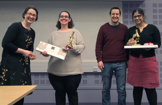
Scientists at the Dunn School spend hours each year using the state-of-the-art microscopes available at the Departmental Facilities, the Central Oxford Structural and Molecular Imaging Centre (COSMIC) and Micron Oxford. Once a year, the Dunn School Bioimaging Facility managers Dr Errin Johnson (Electron Microscopy) and Dr Alan Wainman (Light Microscopy) give users the opportunity to submit their best images for a chance to win one of the highly coveted gold microscope awards.
The 2017 Bioimaging Facility Image Awards were handed out during the Departmental Symposium on 8th January 2018 and were generously supported by the following sponsoring companies: Thermo Scientific, Photometrics, Gatan, Leica Microsystems, Olympus, Zeiss and Microscope Services Ltd. A judging panel formed by Professor Peter Cook, Professor David Greaves and Melissa Wright chose some stunning images from the following winners, amongst over 30 entries:
Light microscopy
Winner: Sonia Muliyil and Clemence Levet (Freeman lab): ‘Gut Instinct’, Muscle surrounding the Drosophila larva gut stained with phalloidin. Imaged on the Zeiss 880 Airyscan.
Runner-up: James Bancroft (Gruneberg lab): ‘Cellular Fireworks’, HeLa cells treated with kinesin inhibitor monastrol stained with tubulin (green), DNA (blue) and expressing GFP-CENP-A (red) to mark the kinetochores. Wide-field fluorescence image.
Electron microscopy
Winner: Rafael Da Silva Custodio (Tang lab): ‘Livin’ on the edge‘, image showing a small group of N. cinerea “climbing” a filopodia tip during the colonisation of human epithelial cells. Acquired on Zeiss Sigma 300 FEG-SEM.
Runner-up: Kenny Moore (James lab): ‘MacroGout’, induced pluripotent stem cell-derived macrophages (red) treated with deoxyguanosine produce crystals of uric acid (cause of Gout) as a by-product of purine catabolism (purple), which protrude from the cell alongside cellular debris (brown). Acquired on the Zeiss Sigma 300 FEG-SEM.
A third category that is gaining popularity each year, is the humorous microscopy –images featuring faces or other curiosities– this year won by Ita Costello (Robertson/Bikoff lab) with ‘This little piggy’; a 3D projection of a day 6 embryoid body, carrying a Blimp1-venus transgene (green) and counterstained with DAPI (blue) and captured on the Olympus FV1000. The runner-up was Alexis Wang (Carvalho lab) with an ‘Emoji yeast’, a S.cerevisiae cell, surprised by our intrusion of privacy, taken on the Tecnai 12 TEM.
To read more about the Dunn School Bioimaging Facility visit: www.dunnschoolbioimaging.co.uk
To find out more about Micron Oxford visit: www.micron.ox.ac.uk
To find out more about cryo-EM at COSMIC visit: http://web.path.ox.ac.uk/~bioimaging/electronmicroscope/cryo_em.html
Written by Anna Caballe (@caballe_anna)