A Whole New World: Dunn School Bioimaging Facility Imaging Awards 2018
The winners of the Dunn School Bioimaging Facility Awards 2018 were announced at the department’s annual Symposium on the 7th of January 2019. This yearly imaging competition hosted by the Dunn School Bioimaging Facility managers, Dr. Errin Johnson (Electron Microscopy) and Dr. Alan Wainman (Light Microscopy), allows researchers to showcase their most beautiful, interesting and inspiring microscopy work taken on one of the many state-of-the-art departmental facility microscopes.
This year’s awards were generously sponsored by Leica Microsystems, Zeiss, Nikon, Olympus, Andor, Photometrics and Agar Scientific. The judging panel, composed of Associate Prof. Omer Dushek, Dr. Sally Cowley and Associate Prof. Monika Gullerova, selected winning and runner-up images in four categories: light microscopy, electron microscopy, humorous microscopy and, a new category, video.
Dr. Wainman said: “2018 has been another great year of imaging in the facility – not least the amazing submissions that were entered in this competition. Many thanks to everyone who participated and congrats to the winners!”
Listed below are the recipients of the 2018 prizes:
Light Microscopy
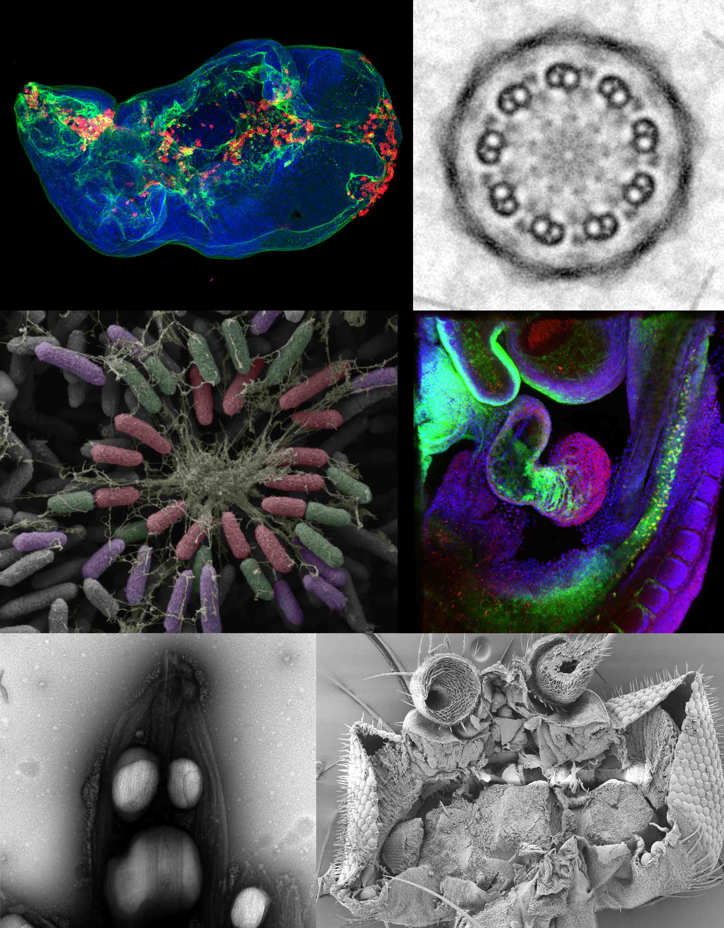
Winner: Dr. Ita Costello (Robertson and Bikoff labs): ‘At the heart of it’. A 3D projection of an early mouse embryo gut tube and developing heart.
Runner-up: Derek Xu (Alberto Baena lab): ‘Cellular Nebula’. Confocal image of a fluorescently labeled Drosophila wing disc.
Electron Microscopy
Winner: Dr. Charlotte Melia (Bharat lab): ‘Gross Fireworks’. A false coloured SEM image of a Pseudomonas aeruginosa biofilm.
Runner-up: Sophia Fochler (Gluenz lab): ‘Microtubule flower’. Composite TEM image a Leishmania axoneme.
Humorous Microscopy
Winner: Dr. Sonia Muliyil and Dr. Clemence Levet (Freeman lab): ‘Cookie monster’. SEM image of an adult Drosophila fly.
Runner-up: Flavia Moreira-Leite (Gull Lab): ‘Screaming Leish’. TEM image of a negatively stained Leishmania parasite.
Video Section (new category for 2018)
Winner: Andriko von Kügelgen (Bharat lab): ‘Crystalised protein’. Cryo-electron tomography and sub-tomogram averaging of crystalised protein filaments.
Runner-up: Dr. Anna Caballe (Raff lab): ‘Mexican wave’. Fluorescently labeled centrosome dividing in an early Drosophila embryo imaged on a confocal microscope.
To read more about the Dunn School Bioimaging Facility see: www.dunnschoolbioimaging.co.uk
Written by Lisa Gartenmann